“`html
Revolutionary Detection of Pancreatic Tumors Inspired by Gila Monster Saliva
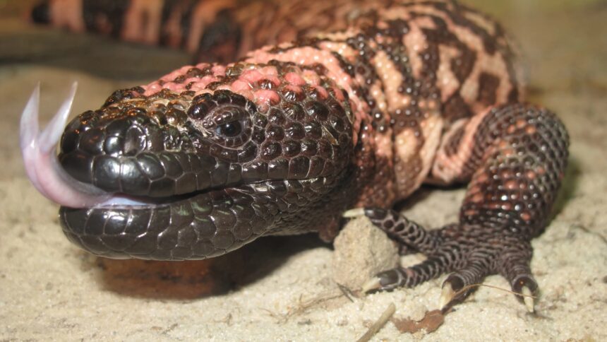
Gila monsters are fascinating reptiles, notable for their unique physical and biochemical characteristics. These lizards, measuring about 1.5 feet in length, are easily identifiable due to their distinctive bumpy scales that exhibit a striking pink and black coloration, along with their stout bodies and characteristic short tails. However, what sets them apart even more is the fact that they belong to one of only two lizard species globally known to produce venom. Although a bite from a Gila monster can be painful and lead to symptoms such as swelling, nausea, and vomiting due to its neurotoxic properties, it is rarely life-threatening. Interestingly, components found in the saliva of these creatures have shown significant potential in aiding the detection of elusive pancreatic tumors.
The Challenge of Insulinomas
Occasionally, beta cells within the pancreas may malfunction and develop small tumors called insulinomas. While these growths are generally benign, they can lead to excessive insulin production which may dangerously lower blood sugar levels—an issue particularly concerning for individuals with diabetes. The resulting hypoglycemia can cause fatigue or even fainting spells. Compounding this challenge is the fact that these tumors often measure less than an inch in diameter, making them difficult for medical professionals to detect accurately. Fortunately, advancements in imaging technology have led to a new variant of PET scans that effectively identifies insulinomas by leveraging the complex chemistry found in Gila monster saliva.
Advancements in Medical Imaging
Prior to this innovative approach inspired by Gila monsters’ saliva, diagnosing patients with insulinoma was an arduous task for healthcare providers; confirming tumor presence could take considerable time.

Credit: Radboud University Medical Center
The Need for Improved Diagnostic Tools
“This condition presents significant challenges,” stated Marti Boss during an interview regarding research published in The Journal of Nuclear Medicine. “While blood tests exist as diagnostic tools—they cannot definitively indicate whether a tumor exists or pinpoint its location.” Various imaging techniques like CT scans or MRIs have been utilized but often fail at detecting insulinomas effectively.
“Historically,” Martin Gotthardt—a nuclear medicine professor involved with this study—added “surgeons would resort to exploratory surgery on patients’ pancreases until they located any existing tumors; if situated at one end it could result in complete removal.” Although living without a pancreas is possible it leads individuals into severe diabetes management challenges requiring constant blood sugar monitoring—highlighting an urgent need for better scanning technologies.
Innovative Solutions Derived from Nature’s Chemistry
Recognizing the potential benefits offered by Gila monster saliva’s chemical composition was pivotal for Gotthardt and Boss’s research team; earlier studies had identified specific compounds within this reptilian secretion capable of binding strongly with GLP1 receptors present on insulinomas’ surfaces—but utilizing raw saliva directly posed stability issues when introduced into human physiology.
To address these concerns researchers synthesized Extendin—a chemically stable version—and combined it with mildly radioactive tracers typically used during standard PET imaging procedures before conducting trials involving 69 adult participants suspected of having insulinoma through Extendin-PET scanning techniques yielding remarkable results: conventional PET methods identified tumors only 65% accurately while Extendin-enhanced scans achieved accuracy rates soaring up towards 95%. Notably among cases where Extendin-PET was paired alongside CT/MRI evaluations revealed additional identification capabilities accounting solely attributable towards this novel method leading ultimately successful surgical removals across all confirmed instances!
[Related:[Related:[Related:[Related:How do bats stay cancer-free? The answer could be lifesaving for humans .
A Bright Future Ahead
Looking ahead researchers aim not only further explore therapeutic applications surrounding Extendin but also work diligently towards integrating modified PET scanning protocols into clinical settings worldwide!
‘We believe our new scanning technique has potential replacing existing methodologies entirely,’ asserted Boss confidently adding ‘…patients who underwent surgeries were completely cured—even those suffering symptoms spanning decades.’
Source
“`


